

商品详情
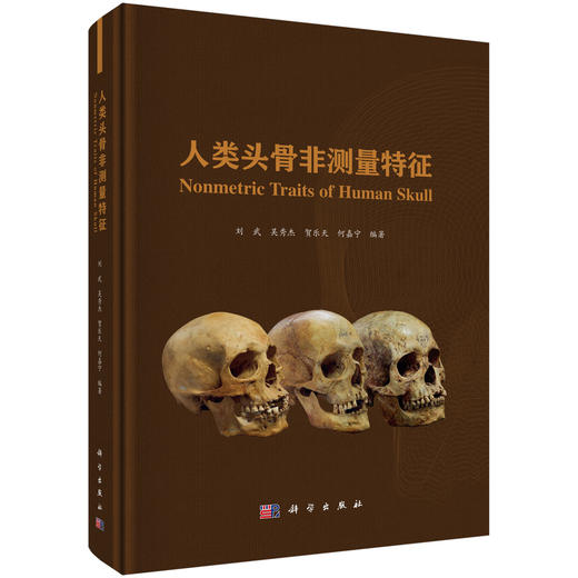
书名:人类头骨非测量特征
定价:498.0
ISBN:9787030856951
作者:刘武等
版次:1
出版时间:2026-07
内容提要:
目录:
目录
序一 Foreword 1 i
序二 Foreword 2 iii
前言 Preface v
Contents
第一章 引言
Chapter One Introduction
1.1 人类头骨非测量特征定义与分类 2
Definition and classifications of the nonmetric traits of the human skull
1.2 非测量特征研究历史 6
History of the nonmetric trait studies
1.3 非测量特征的影响控制机制 8
The influence to the nonmetric traits
1.4 非测量特征的研究价值 14
The research values of the nonmetric traits
1.5 本书写作原则与导读 17
Guide to use the book
第二章 头盖部非测量特征
Chapter Two Nonmetric Traits of the Calvaria
2.1 颅骨轮廓形状(颅形)Cranial form 21
2.2 额中缝 Metopic suture, Metopism 25
2.3 鼻上缝 Supranasal suture (SPS) 29
人类头骨非测量特征 Nonmetric T viii raits of Human Skull
2.4 眶上神经沟 Supraorbital nerve groove 32
额沟 Frontal groove, Frontal sulcus
2.5 矢状脊 Sagittal keeling 34
2.6 顶孔 Parietal foramen 37
2.7 前囟后部凹陷 Postbregmatic depression (PBD) 39
2.8 顶间沟 Interparietal groove, Interparietal sulcus 41
2.9 顶孔至人字区平坦 Lambdoid flat 43
顶孔至人字区凹陷 Lambdoid depression
2.10 枕区突隆 Occipital bunning 46
2.11 翼区连接方式 Sutural variation at pterion 48
2.12 角圆枕 Angular torus 50
2.13 外耳道骨肿 External auditory exostosis 52
耳圆枕 Auditory torus
2.14 耳孔上凹 Suprameatal depression 54
耳孔上棘 Suprameatal spine
2.15 乳突大小 Mastoid process 56
2.16 乳突上脊 Supramastoid crest 58
2.17 乳突缝 Squamomastoid suture 60
2.18 乳突旁隆起 Juxtamastoid eminence, Paramastoid eminence 62
2.19 乳突切迹 Mastoid notch 64
二腹肌沟 Digastric groove
2.20 枕乳脊 Occipitomastoid crest 66
2.21 乳突孔 Mastoid foramen 67
2.22 乳突后隆起 Retromastoid process, Retromastoid tubercle 69
2.23 顶颞缝 Parietotemporal suture 71
2.24 颞线 Temporal line 72
2.25 枕外隆凸 External occipital protuberance 75
2.26 最上项线 Highest nuchal line, Supreme nuchal line 77
2.27 枕圆枕 Occipital torus 79
2.28 枕外脊 External occipital crest 82
2.29 枕项平面转折 Transition from the occipital plane to the nuchal plane 84
2.30 枕孔 Occipital foramen 86
2.31 枕横缝 Mendosal suture 88
星点间缝 Biasterionic suture
2.32 枕外隆凸上窝 Suprainiac fossa 90
枕外隆凸上凹陷 Suprainiac depression
2.33 茎突 Styloid process 92
2.34 岩鳞导管 Petrosquamous emissaries 94
2.35 颞骨鼓板粗壮度 Tympanic plate of the temporal bone 96
第三章 面部非测量特征
Chapter Three Nonmetric Traits on the Facial Bones
3.1 眼眶形状 Orbital shape (OBS) 99
3.2 眶上区形态变异 Morphological variations on the supraorbital region 104
3.3 眶上孔 Supraorbital foramen 110
眶上切迹 Supraorbital notch
3.4 眼眶外下缘 Inferolateral margin of the orbit 113
眼眶外下缘圆钝 Rounding of the inferolateral margin of the orbit
3.5 眶下孔 Infraorbital foramen 115
3.6 眶下缝 Infraorbital suture 118
3.7 鼻额缝与额颌缝的走向 Course of frontonasal suture and frontomaxillary suture 120
鼻额缝走向 Course of nasofrontal suture (NFS)
3.8 鼻梁冠状隆起 Profile of the nasal saddle and nasal roof 124
鼻骨轮廓 Nasal bone contour (NBC)
3.9 眉间至鼻根点凹陷 Profile of the infraglabellar notch 128
3.10 鼻骨形状 Shape of the nasal bones, Nasal bone shape (NBS) 130
3.11 梨状孔形状 Nasal aperture shape (NAS), pyriforme orifice shape 133
3.12 梨状孔下缘形态 135
Inferior margin of the nasal aperture, Inferior nasal aperture (INA)
3.13 鼻下窝 Subnasal fossa 141
3.14 鼻前棘 Anterior nasal spine (ANS) 143
3.15 鼻骨孔 Nasal foramen 146
3.16 颧三角 Zygomatic trigone 148
3.17 颧结节 Zygomatic (malar) tuberosity 150
3.18 颧颌缝下缘结节 152
Zygomaxillary tubercle, Malar tubercle (MT), Malar process
3.19 颧上颌缝 Zygomaxillary suture (ZS) 155
3.20 颧颌缝脊 Ridge on the zygomaxillary suture 157
3.21 颧横缝痕迹 Transverse zygomatic suture vestige 158
二分颧骨 Divided zygoma (DZ), Os zygomaticum bipartitum,Bipartite zygomatic bone
日本骨 Os japonicum
人类头骨非测量特征 Nonmetric T x raits of Human Skull
3.22 颧面孔 Zygomatico-facial foramen 163
3.23 犬齿窝 Canine fossa 165
3.24 上颌骨颧突下缘形态 Lower margin of cheek bones 167
颧切迹 Malar incisure
3.25 梨状孔外侧面隆凸 Bulging between orbit and pyriforme orifice 168
3.26 颧骨后结节 Posterior zygomatic tubercle (PZT) 170
颧骨缘突 Marginal process, Marginal tubercle
3.27 鼻骨前下端突起 Projection of the anterior extremity of the nasal bones 172
第四章 颅底部非测量特征
Chapter Four Nonmetric Traits on the Cranial Basal View
4.1 腭形 Palate shape (PS) 175
齿弓轮廓 Contour of the dental arcade and palate
4.2 门齿孔 Incisive foramen 178
4.3 腭圆枕 Palatine torus 180
4.4 上颌圆枕 Maxillary torus 183
4.5 腭横缝 Transverse palatine suture (TPS), Transverse palatomaxillary suture 185
4.6 腭骨桥 Palatine bridging 189
4.7 副腭小孔 Accessory lesser palatine foramen 191
腭小孔 Lesser palatine foramen
4.8 鞍桥 Clinoid bridging, Clino-clinoid bridge, Sella bridges, Ponticuli sellae 194
4.9 翼棘桥 Pterygospinous bridge 196
翼棘孔 Pterygospinous foramen
斯维尼孔 Foramen of Civinini
赫依特孔 Foramen of Hyrtl
4.10 颅咽管 Craniopharyngeal canal 199
4.11 咽部窝 Pharyngeal fossa 201
4.12 咽部突(咽结节)Pharyngeal tubercle 203
4.13 卵圆孔–棘孔变异 Variants of oval foramen - spinous foramen 205
4.14 鼓板开裂 Tympanic dehiscence 210
胡施克孔 Foramen of Huschke
4.15 舌下神经管骨桥 Hypoglossal canal bridged 213
二分舌下神经管 Hypoglossal canal double
4.16 颈动脉孔外口 External opening of the carotid canal 216
鼓小管下口 Apertura inferior canaliculi tympanici
4.17 颈静脉孔骨桥 218
Bony bridging of jugular foramen, Jugular foramen bridging
4.18 髁前结节 Precondylar tubercle or process 221
4.19 髁旁突 Paracondylar process 224
4.20 第三髁 Third occipital condyle 226
枕–枢椎关节面 Condylus tertius, Odonto-occipital articulation
4.21 基底中央孔(基底中央管)Median basilar canal 228
4.22 二分枕髁关节面 Condylar facet double, Double condylar facet 230
4.23 髁后管(髁后孔) 232
Condylar canal (foramen), Postcondylar canal, Posterior condylar canal
4.24 髁间管 Lateral condylar canal 234
髁中间管 Intermediate condylar canal
髁旁孔 Paracondylar foramen
4.25 枕骨大孔形状 Shape of the foramen magnum 236
4.26 静脉窦系统 Venous sinuses system 238
第五章 缝间骨
Chapter Five Sutural Bones
5.1 前囟骨 Bregmatic bone 243
前囟小骨 Ossicle at bregma, Fontanelle ossicle
5.2 冠状缝小骨 Coronal ossicle 246
5.3 矢状缝间骨 Sagittal suture bone 248
矢状缝小骨 Sagittal suture ossicle
5.4 人字点骨 Apical bone 251
人字点小骨 Ossicle at lambda, Os apicis
5.5 人字缝间骨 Lambdoid bone 254
人字缝小骨 Lambdoid ossicle
5.6 印加骨 Inca bone, Os Incae 256
5.7 顶切迹骨 Parietal notch bone 261
5.8 星点骨 Asterionic bone 264
星点小骨 Ossicle at asterion
5.9 枕乳缝间骨 Occipitomastoid ossicle, Occipitomastoid wormians 267
5.10 翼上骨 Epiteric bone 270
5.11 颞鳞骨 Squamous bone 273
颞鳞小骨 Squamous ossicle, Temporo-squamous ossicle (TESO)
人类头骨非测量特征 Nonmetric T xii raits of Human Skull
5.12 额中小骨 Metopic ossicle, Metopic fontanelle 275
额中裂缝 Metopic fissure
第六章 下颌骨非测量特征
Chapter Six Nonmetric Traits of the Mandible
6.1 下颌联合颊侧面 Buccal Aspect of the Mandibular Symphysis 279
6.1-1 中央脊 Central keel 280
6.1-2 联合结节 Symphyseal tubercle, Tuber symphysis 282
6.1-3 侧结节 Lateral tubercle 283
6.1-4 颏窝 Mental fossa 284
6.1-5 下颌联合前部弯… Anterior symphyseal incurvature 286
6.1-6 下颌联合前部倾角 Anterior symphyseal angle 287
6.1-7 颏隆凸 Mentum osseum 288
下巴 Chin
6.1-8 颏下切迹 Submental incisure 292
6.2 下颌联合舌侧面 Lingual Aspect of the Mandibular Symphysis 293
6.2-1 齿槽平台 Alveolar planum 294
6.2-2 上横圆枕 Superior transverse torus 296
6.2-3 下横圆枕 Inferior transverse torus 297
6.2-4 颏舌窝 Genioglossal fossa 298
6.2-5 二腹肌窝 Digastric fossa 299
6.2-6 齿槽颏孔 Alveolar mental foramen 300
副齿槽颏孔 Accessory alveolar foramina
6.2-7 下颌联合舌侧面中央纵沟 Median groove 302
下颌联合舌侧面旁矢状纵沟 Parasagittal groove
6.2-8 颏结节 Genial tubercle 304
颏棘 Mental spine
6.2-9 颏舌孔 Genial foramina, Genioglossal foramen 307
6.3 下颌体颊侧面 Buccal Aspect of the Mandibular Corpus 309
6.3-1 侧隆起 Lateral prominence 310
6.3-2 上侧圆枕 Lateral superior torus 312
6.3-3 边缘圆枕 Marginal torus 313
6.3-4 圆枕间沟 Intertoral sulcus 314
6.3-5 前边缘结节 Anterior marginal tubercle 315
6.3-6 后边缘结节 Posterior marginal tubercle 316
6.3-7 颏孔位置与数量 Number and position of mental foramen 317
6.3-8 摇椅型下颌 Rocker jaw 319
6.3-9 臼齿外沟 Extramolar sulcus 322
6.3-10 臼齿后空间 Retromolar space 323
6.4 下颌体舌侧面 Lingual Aspect of the Mandibular Corpus 325
6.4-1 下颌圆枕 Mandibular torus 326
6.4-2 下颌下窝 Submandibular fossa 329
6.4-3 舌骨肌线 Mylohyoid line 330
6.4-4 齿槽隆凸 Alveolar prominence 331
6.5 下颌支外侧面 External Aspect of the Mandibular Ramus 332
6.5-1 下颌切迹对称性 Mandibular notch symmetry 333
6.5-2 髁突相对冠突高度 Condyle height relative to the coronoid 335
6.5-3 下颌切迹相对髁突位置 336
Position of the mandibular notch relative to the condyle
6.5-4 髁突外侧脊 Ectocondyloid crest (crista) 337
6.5-5 髁突颊侧下结节 Lateral subcondyloid tubercle 338
6.5-6 下颌支侧隆起 Lateral ramus eminence 339
6.5-7 髁突下窝 Subcondylar fossa 340
6.5-8 咬肌窝 Masseteric fossa 341
6.5-9 下颌角后缘形态 Posterior gonial morphology 342
下颌角外翻 Gonial eversion
6.5-10 下颌角前切迹 Antegonial notch, Preangular mandibular notch 343
6.6 下颌支内侧面 Medial Aspect of the Mandibular Ramus 344
6.6-1 冠突内脊 Endocoronoid crest 345
6.6-2 髁突内脊 Endocondyloid crest 347
6.6-3 冠突髁突内脊间三角平台(窝) 348
Triangular planum, Triangular fossa
6.6-4 翼肌上结节 349
Superior pterygoid tubercle, Tuberculum pterygoideum superius (tps)
6.6-5 翼肌下结节 350
Tuberculum pterygoideum inferius (tpi), Inferior pterygoid tubercle
6.6-6 翼内肌结节 Medial pterygoid tubercle (MPT) 351
翼内肌上结节 Superior medial pterygoid tubercle
6.6-7 翼肌粗隆 Pterygoid tuberosoties 353
6.6-8 翼肌窝 Pterygoid fossa 354
6.6-9 下颌孔 Mandibular foramen 355
6.6-10 副下颌孔 Accessory mandibular foramen 356
人类头骨非测量特征 Nonmetric T xiv raits of Human Skull
6.6-11 下颌舌骨沟 Mylohyoid groove 357
下颌舌骨沟桥 Mylohyoid bridging
6.6-12 臼齿后孔 Retromolar foramen 360
罗宾逊孔 Robosin canal
第七章 非测量特征采集与数据分析方法
Chapter Seven Data Collection and Analysis Methods of the Nonmetric Traits
7.1 观测标本的选择 Selection of the specimens 362
7.2 非测量特征的选择 Selection of the nonmetric traits 363
7.3 非测量特征数据的采集 Nonmetric traits data collection 364
7.4 观察者误差的评估 Evaluation of the observer’s errors 365
7.5 数据初步分析 Preliminary data analysis 366
7.6 人群关系分析 Analysis of population relationship 370
7.7 序列等级特征有/无判断的参考标准 373
Reference criteria of nonmetric traits assessment
第八章 非测量特征在研究中的应用
Chapter Eight Applications of Nonmetric Traits
8.1 人类演化 Human evolution 380
8.2 人群关系及微观演化 Population relationships and microevolution 381
8.3 功能及环境适应 Functional and environmental adaptation 384
8.4 个体鉴定与识别 Individual identification 386
参考文献 References 387
索引 Subject Index 405
后记 Postscript 415
图2-Ⅰ 颅骨前面观(A)、后面观(B)和顶面观(C), 20
展示部分头盖部非测量特征
Figure 2-Ⅰ Anterior (A), posterior (B) and superior views (C) of calvaria
showing some cranial nonmetric traits
图2-Ⅱ 颅骨侧面观,展示部分头盖部非测量特征 20
Figure 2-Ⅱ Lateral view of a cranium showing some cranial nonmetric traits
图2.1-1 颅骨前面观,展示颅顶外轮廓形状 21
Figure 2.1-1 Anterior view of the crania showing the contour shape at the top of the crania
图2.1-2 颅骨后面观,展示侧壁外轮廓形状 21
Figure 2.1-2 Posterior view of the crania showing the contour shape at the lateral walls
图2.1-3 颅骨顶面观,展示颅顶面外轮廓形状 22
Figure 2.1-3 The contour shape of the crania in superior view
图2.2-1 颅骨前面观,展示额中缝类型 25
Figure 2.2-1 Anterior view of the crania showing the expression types of the metopic suture
图2.2-2 额中缝的形态 26
Figure 2.2-2 The shapes of the metopic suture
图2.2-3 部分额中缝的三种表现类型 26
Figure 2.2-3 Three types of the partial persistence of the metopic suture
图2.3-1 鼻上缝(A)和额中缝(B) 29
Figure 2.3-1 Supranasal suture (A) and metopic suture (B)
图2.3-2 鼻上缝的形状变异 30
Figure 2.3-2 The variants of the supranasal suture shape
图2.3-3 鼻上缝类型(根据愈合程度) 30
Figure 2.3-3 The types of supranasal suture according to the degree of persistence
插图目录
List of Figures
人类头骨非测量特征 Nonmetric T xvi raits of Human Skull
图2.4 眶上神经沟的不同表现 32
Figure 2.4 Different expressions of supraorbital nerve grooves
图2.5-1 颅骨顶侧面观,展示额矢状脊和顶矢状脊 34
Figure 2.5-1 Superior-lateral view of the skull showing the frontal and sagittal keelings
图2.5-2 矢状脊表现形式 35
Figure 2.5-2 Different expressions of sagittal keeling
图2.6 顶孔的变异 37
Figure 2.6 The variants of the parietal foramen
图2.7 前囟后部凹陷的表现 39
Figure 2.7 The expressions of postbregmatic depression
图2.8-1 顶间沟类型,按照发育程度 41
Figure 2.8-1 Types of interparietal groove according development
图2.8-2 顶间沟类型,按照出现位置 42
Figure 2.8-2 Types of interparietal groove according positions
图2.9-1 顶孔至人字区的形态变异 43
Figure 2.9-1 The morphology variants from the parietal foramen to the lambda region
图2.9-2 顶孔至人字区平坦不同表现 44
Figure 2.9-2 The variants of lambdoid flat
图2.10 颅骨左侧观,展示枕区突隆 46
Figure 2.10 Lateral view of the crania showing the occipital bunning
图2.11 翼区连接方式 48
Figure 2.11 The articulation variants of the pterion region
图2.12 角圆枕的变异 50
Figure 2.12 The variants of angular torus
图2.13-1 外耳道骨肿的变异 52
Figure 2.13-1 The variants of external auditory exostosis
图2.13-2 外耳道骨肿的表现 53
Figure 2.13-2 The expression of external auditory exostosis
图2.14-1 耳孔上凹(A)、耳孔上棘(B)及两者都存在(C) 54
Figure 2.14-1 Suprameatal depression (A), suprameatal spine (B) and both present (C)
图2.14-2 耳孔上棘与耳孔上凹的变异 55
Figure 2.14-2 The variants of the suprameatal depression and suprameatal spine
图2.15 乳突大小变异 56
Figure 2.15 The size variants of mastoid process
图2.16 乳突上脊的变异 58
插图目录 List of Figures xvii
Figure 2.16 The variants of the supramastoid crest
图2.17-1 乳突缝的变异 60
Figure 2.17-1 The variants of squamomastoid suture
图2.17-2 乳突缝的不同表现 61
Figure 2.17-2 Degrees of persistence of a squamomastoid suture
图2.18-1 颅骨底部观,展示乳突后部的三个特征 62
Figure 2.18-1 Basal view of the cranium showing the three traits posterior
to the mastoid process
图2.18-2 乳突旁隆起的变异 63
Figure 2.18-2 The variants of the juxtamastoid eminence
图2.19 乳突切迹的变异 64
Figure 2.19 The variants of mastoid notch
图2.20 枕乳脊的变异 66
Figure 2.20 The variants of occipitomastoid crest
图2.21 乳突孔的变异 68
Figure 2.21 The variants of mastoid foramen
图2.22-1 颅骨底面观,展示乳突后隆起在项平面上的位置 69
Figure 2.22-1 Basal view of the cranium showing the retromastoid process on the nuchal plane
图2.22-2 乳突后隆起的表现 70
Figure 2.22-2 The expression of retromastoid process
图2.23 颅骨侧面观,展示顶颞缝的变异 71
Figure 2.23 Lateral view of the crania showing the variants of parietotemporal suture
图2.24-1 颅骨侧面观,展示颞线的走向 72
Figure 2.24-1 Lateral view of the crania showing the extension direction of the temporal line
图2.24-2 颅骨侧面观,展示两颞线的宽度 72
Figure 2.24-2 Lateral view of the crania showing the width between the two temporal lines
图2.24-3 颅骨侧面观,展示颞线的发育程度 73
Figure 2.24-3 Lateral view of the crania showing the development degree of the temporal line
图2.24-4 颅骨侧面观,展示颞线的粗糙度 73
Figure 2.24-4 Lateral view of the crania showing the roughness of the temporal line
图2.24-5 颅骨后侧面观,展示颞线末端的位置 74
人类头骨非测量特征 Nonmetric T xviii raits of Human Skull
Figure 2.24-5 Posterolateral view of the crania showing the position of the end of the temporal line
图2.25 枕骨侧面观,展示枕外隆凸的变异 75
Figure 2.25 Lateral view of the occipital bone showing the variants of external occipital protuberance
图2.26 枕骨后面观,展示最上项线的变异 77
Figure 2.26 Posterior view of the occipital bone showing the variants of the highest nuchal line
图2.27 枕骨后面观,展示枕圆枕的变异 79
Figure 2.27 Posterior view of the occipital bone showing the variants of occipital torus
图2.28 枕骨后面观,展示枕外脊的变异 82
Figure 2.28 Posterior view of the occipital bone showing the variants of external
occipital crest
图2.29 枕骨侧面观,展示枕项平面转折的变异 84
Figure 2.29 Lateral view of the occipital bone showing the variants of transition from occipital to nuchal plane
图2.30 枕骨大孔前缘观,展示枕孔的变异 86
Figure 2.30 The region anterior to the foramen magnum showing the variants of occipital foramen
图2.31-1 颅骨后面观图绘,展示枕横缝的位置变异 88
Figure 2.31-1 The posterior view of the crania drawing showing the variants of mendosal suture
图2.31-2 枕骨左侧面观,展示枕横缝的变异 89
Figure 2.31-2 Left lateral view of the occipital bones showing the variants of mendosal suture
图2.32-1 更新世古人类枕骨,展示枕外隆凸上窝的表现 90
Figure 2.32-1 Occipital bones of the Pleistocene hominins showing the expression
of the suprainiac fossa
图2.32-2 现代人颅骨后面观,展示枕外隆凸上凹陷的变异 91
Figure 2.32-2 Posterior view of the modern human crania showing the variants of suprainiac depression
图2.33-1 颅骨底面观,展示茎突位置 92
Figure 2.33-1 Basal view of the cranium showing the position of styloid process
图2.33-2 颅骨底面观,展示枕骨大孔左侧茎突的变异 93
Figure 2.33-2 Basal views of the crania showing the variants of styloid process
on the left side of foramen magnum
图2.34 岩鳞导管的不同表现 94
Figure 2.34 Different expressions of the petrosquamous emissaries
图2.35 颞骨鼓板发育情况 96
Figure 2.35 The development of the tympanic plate of the temporal bone
图3-Ⅰ 颅骨前面观,展示部分面部非测量特征 98
Figure 3-Ⅰ Anterior view of the cranium showing some nonmetric traits on the facial bones
图3-Ⅱ 颅骨前侧面观,展示部分面部非测量特征 98
Figure 3-Ⅱ Anterolateral view of the cranium showing some nonmetric traits on the facial bones
图3.1-1 眼眶形状的四种类型 99
Figure 3.1-1 The four categories of the orbital shape
图3.1-2 根据上眶缘、上侧角和下眶缘的走向计数的眼眶形状的方法 100
Figure 3.1-2 Scoring of orbital shape through the relative inclination of the superior,
lateral and inferior orbital elements
图3.1-3 根据眼眶边缘形态对眼眶形状分类的三个类型 100
Figure 3.1-3 The three categories of orbital shape through the orbital margin
图3.1-4 眼眶形状分类的六个类型 101
Figure 3.1-4 The six categories of the orbital shape
图3.2-1 眶上区域的一些非测量性状 104
Figure 3.2-1 Some nonmetric traits on the supraorbital region
图3.2-2 眉脊发育程度的五种类型 105
Figure 3.2-2 The five categories of the development degree of supraorbital ridges
图3.3-1 眶上孔和眶上切迹 110
Figure 3.3-1 Supraorbital foramen and supraorbital notch
图3.3-2 眶上孔(眶上切迹)位置的变异 111
Figure 3.3-2 The variants in the position of supraorbital foramen or supraorbital notch
图3.3-3 眶上孔(眶上切迹)数量和形态的变异 111
Figure 3.3-3 The variants in the number and morphology of supraorbital foramen or
supraorbital notch
图3.4 眼眶外下缘圆钝的三个类型 113
Figure 3.4 The three categories in the development degree of inferolateral margin of the orbit
图3.5-1 眶下孔不同表现类型 115
人类头骨非测量特征 Nonmetric T xx raits of Human Skull
Figure 3.5-1 Different expressions of the infraorbital foramen
图3.5-2 眶下孔的变异 116
Figure 3.5-2 The variants of the infraorbital foramen
图3.6-1 眶下缝的三种类型 118
Figure 3.6-1 Diagram showing the three categories of the infraorbital suture
图3.6-2 眶下缝的变异 119
Figure 3.6-2 The variants of the infraorbital suture
图3.7-1 额鼻缝与额颌缝的走向变异 120
Figure 3.7-1 The variants in the course of frontonasal and frontomaxillary sutures
图3.7-2 额鼻缝和额颌缝的高度变异 121
Figure 3.7-2 The variants in the height of frontonasal and frontomaxillary suture
图3.7-3 额鼻缝和额颌缝相交的形态 121
Figure 3.7-3 The variants of the intersection between frontonasal and frontomaxillary suture
图3.7-4 鼻额缝的四个类型 122
Figure 3.7-4 The four categories of the shape of nasofrontal suture
图3.8-1 鼻梁冠状隆起的不同表现 124
Figure 3.8-1 The variants of profile of the nasal saddle and nasal roof
图3.8-2 鼻骨轮廓的分类级别 125
Figure 3.8-2 Diagrammatic representations showing the different states of the nasal bone contour
图3.9 眉间至鼻根点凹陷 128
Figure 3.9 Profile of the infraglabellar notch
图3.10-1 鼻骨形状的分类 130
Figure 3.10-1 The types of nasal bone shape
图3.10-2 鼻骨形状的分类 131
Figure 3.10-2 Photographic exemplars of the different types of nasal bone shape
图3.11 梨状孔形状的分类 133
Figure 3.11 Diagrammatic representations and variants of the nasal aperture shape
图3.12-1 梨状孔下缘形态变异 135
Figure 3.12-1 The variants of morphology of inferior nasal aperture
图3.12-2 图示梨状孔下缘形态的分类 136
Figure 3.12-2 Diagrammatic representation of the variants of inferior nasal aperture
图3.12-3 梨状孔下缘形态结构 137
Figure 3.12-3 The anatomic features of the inferior margin of the nasal aperture
图3.12-4 梨状孔下缘的形态变异 138
插图目录 List of Figures xxi
Figure 3.12-4 The variants of the inferior margin of the nasal aperture
图3.13 鼻下窝不同类型 141
Figure 3.13 The variants of the subnasal fossa
图3.14 鼻前棘的变异 143
Figure 3.14 Diagrammatic representations and variants of the anterior nasal spine
图3.15-1 鼻骨孔数量的变异 146
Figure 3.15-1 The variants in the number of nasal foramina
图3.15-2 鼻骨孔位置的变异 147
Figure 3.15-2 The variants in the position of nasal foramina
图3.15-3 鼻骨孔大小的变异 147
Figure 3.15-3 The variants in the size of nasal foramen
图3.16 颧三角的分级 148
Figure 3.16 The classification types of the zygomatic trigone
图3.17 颧结节的四种类型 150
Figure 3.17 The four types of zygomatic tuberosity
图3.18-1 颧颌缝下缘结节的分级类型 152
Figure 3.18-1 Diagrammatic representation of different character states of zygomaxillary tubercle
图3.18-2 颧颌缝下缘结节的分级类型 153
Figure 3.18-2 The different character states of zygomaxillary tubercle
图3.19 颧上颌缝的分类 155
Figure 3.19 The character states of zygomaxillary suture
图3.20 颧颌缝脊的三个类型 157
Figure 3.20 The three categories of the ridge on the zygomaxillary suture
图3.21-1 分隔颧骨的四种类型 159
Figure 3.21-1 The four categories of transverse zygomatic suture vestige
图3.21-2 图示颧横缝痕迹的各种变异 159
Figure 3.21-2 Graphical representation the variants of the transverse zygomatic suture vestige
图3.21-3 图示完全类型分隔颧骨的各种表现 160
Figure 3.21-3 The variants of complete sutural division of the zygomatic bone
图3.21-4 仅仅出现在颧骨内面的二分颧骨 162
Figure 3.21-4 Divided zygoma only occurs at inner surface of the zygomatic bone
图3.22-1 颧面孔的不同表现类型 163
Figure 3.22-1 The variants of the zygomatico-facial foramen
图3.22-2 颧面孔位置的变异 164
人类头骨非测量特征 Nonmetric T xxii raits of Human Skull
Figure 3.22-2 The variants in the position of the zygomaticofacial foramina
图3.23 犬齿窝的不同表现 165
Figure 3.23 The variants of the canine fossa
图3.24 上颌骨颧突下缘形态的变异 167
Figure 3.24 The variants in the lower margin of cheek bones
图3.25 梨状孔外侧面隆凸的变异 168
Figure 3.25 The variations of the bulging between orbit and pyriforme orifice
图3.26 颧骨后结节突起的不同类型 170
Figure 3.26 The variants of posterior zygomatic tubercle
图3.27 鼻骨前下端突起不同类型 172
Figure 3.27 The types of projection of the anterior extremity of the nasal bones
图4-Ⅰ 颅骨底面观部分非测量特征 174
Figure 4-Ⅰ Basal view of cranium showing some nonmetric traits
图4-Ⅱ 颅骨内面观部分非测量特征 174
Figure 4-Ⅱ Internal view of cranium showing some nonmetric traits
图4.1-1 齿弓形状的三个类型 175
Figure 4.1-1 Three character states of contour of the dental arcade and palate
图4.1-2 腭形(齿弓轮廓)的四个类型 176
Figure 4.1-2 Four character states of contour of the dental arcade and palate
图4.2-1 硬腭面展示门齿孔(A)及上颌骨切面展示门齿管(B) 178
Figure 4.21-1 The palatine showing the incisive foramen (A) and incisive canal (B)
图4.2-2 门齿孔的不同类型 179
Figure 4.2-2 The variants of the incisive foramens
图4.3 腭圆枕的四个类型 180
Figure 4.3 The four categories of the palatine torus
图4.4-1 上颌骨底面和侧面观,展示上颌圆枕 183
Figure 4.4-1 Basal and lateral views of the maxilla showing the maxillary torus
定价:498.0
ISBN:9787030856951
作者:刘武等
版次:1
出版时间:2026-07
内容提要:
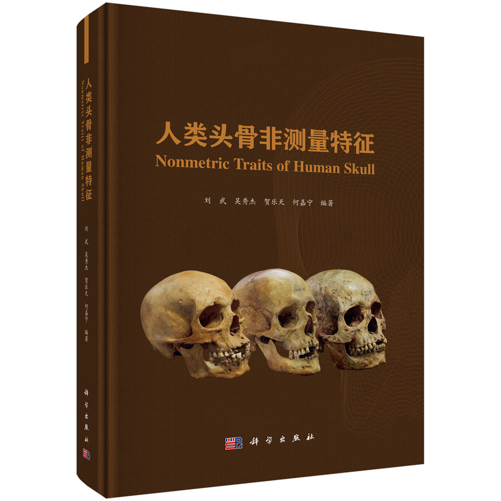
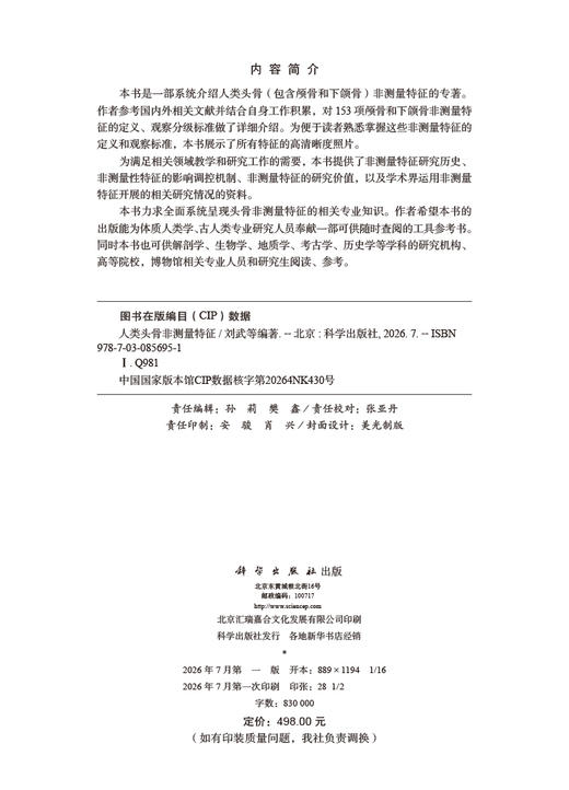
本书是一部系统介绍人类头骨(包含颅骨和下颌骨)非测量特征的专著。作者参考国内外相关文献并结合自身工作积累,对153项颅骨和下颌骨非测量特征的定义、观察分级标准做了详细介绍。为便于读者熟悉掌握这些非测量特征的定义和观察标准,本书展示了所有特征的高清晰度照片。
为满足相关领域教学和研究工作的需要,本书提供了非测量特征研究历史、非测量性特征的影响调控机制、非测量特征的研究价值,以及学术界运用非测量特征开展的相关研究情况的资料。
目录:
目录
序一 Foreword 1 i
序二 Foreword 2 iii
前言 Preface v
Contents
第一章 引言
Chapter One Introduction
1.1 人类头骨非测量特征定义与分类 2
Definition and classifications of the nonmetric traits of the human skull
1.2 非测量特征研究历史 6
History of the nonmetric trait studies
1.3 非测量特征的影响控制机制 8
The influence to the nonmetric traits
1.4 非测量特征的研究价值 14
The research values of the nonmetric traits
1.5 本书写作原则与导读 17
Guide to use the book
第二章 头盖部非测量特征
Chapter Two Nonmetric Traits of the Calvaria
2.1 颅骨轮廓形状(颅形)Cranial form 21
2.2 额中缝 Metopic suture, Metopism 25
2.3 鼻上缝 Supranasal suture (SPS) 29
人类头骨非测量特征 Nonmetric T viii raits of Human Skull
2.4 眶上神经沟 Supraorbital nerve groove 32
额沟 Frontal groove, Frontal sulcus
2.5 矢状脊 Sagittal keeling 34
2.6 顶孔 Parietal foramen 37
2.7 前囟后部凹陷 Postbregmatic depression (PBD) 39
2.8 顶间沟 Interparietal groove, Interparietal sulcus 41
2.9 顶孔至人字区平坦 Lambdoid flat 43
顶孔至人字区凹陷 Lambdoid depression
2.10 枕区突隆 Occipital bunning 46
2.11 翼区连接方式 Sutural variation at pterion 48
2.12 角圆枕 Angular torus 50
2.13 外耳道骨肿 External auditory exostosis 52
耳圆枕 Auditory torus
2.14 耳孔上凹 Suprameatal depression 54
耳孔上棘 Suprameatal spine
2.15 乳突大小 Mastoid process 56
2.16 乳突上脊 Supramastoid crest 58
2.17 乳突缝 Squamomastoid suture 60
2.18 乳突旁隆起 Juxtamastoid eminence, Paramastoid eminence 62
2.19 乳突切迹 Mastoid notch 64
二腹肌沟 Digastric groove
2.20 枕乳脊 Occipitomastoid crest 66
2.21 乳突孔 Mastoid foramen 67
2.22 乳突后隆起 Retromastoid process, Retromastoid tubercle 69
2.23 顶颞缝 Parietotemporal suture 71
2.24 颞线 Temporal line 72
2.25 枕外隆凸 External occipital protuberance 75
2.26 最上项线 Highest nuchal line, Supreme nuchal line 77
2.27 枕圆枕 Occipital torus 79
2.28 枕外脊 External occipital crest 82
2.29 枕项平面转折 Transition from the occipital plane to the nuchal plane 84
2.30 枕孔 Occipital foramen 86
2.31 枕横缝 Mendosal suture 88
星点间缝 Biasterionic suture
2.32 枕外隆凸上窝 Suprainiac fossa 90
枕外隆凸上凹陷 Suprainiac depression
2.33 茎突 Styloid process 92
2.34 岩鳞导管 Petrosquamous emissaries 94
2.35 颞骨鼓板粗壮度 Tympanic plate of the temporal bone 96
第三章 面部非测量特征
Chapter Three Nonmetric Traits on the Facial Bones
3.1 眼眶形状 Orbital shape (OBS) 99
3.2 眶上区形态变异 Morphological variations on the supraorbital region 104
3.3 眶上孔 Supraorbital foramen 110
眶上切迹 Supraorbital notch
3.4 眼眶外下缘 Inferolateral margin of the orbit 113
眼眶外下缘圆钝 Rounding of the inferolateral margin of the orbit
3.5 眶下孔 Infraorbital foramen 115
3.6 眶下缝 Infraorbital suture 118
3.7 鼻额缝与额颌缝的走向 Course of frontonasal suture and frontomaxillary suture 120
鼻额缝走向 Course of nasofrontal suture (NFS)
3.8 鼻梁冠状隆起 Profile of the nasal saddle and nasal roof 124
鼻骨轮廓 Nasal bone contour (NBC)
3.9 眉间至鼻根点凹陷 Profile of the infraglabellar notch 128
3.10 鼻骨形状 Shape of the nasal bones, Nasal bone shape (NBS) 130
3.11 梨状孔形状 Nasal aperture shape (NAS), pyriforme orifice shape 133
3.12 梨状孔下缘形态 135
Inferior margin of the nasal aperture, Inferior nasal aperture (INA)
3.13 鼻下窝 Subnasal fossa 141
3.14 鼻前棘 Anterior nasal spine (ANS) 143
3.15 鼻骨孔 Nasal foramen 146
3.16 颧三角 Zygomatic trigone 148
3.17 颧结节 Zygomatic (malar) tuberosity 150
3.18 颧颌缝下缘结节 152
Zygomaxillary tubercle, Malar tubercle (MT), Malar process
3.19 颧上颌缝 Zygomaxillary suture (ZS) 155
3.20 颧颌缝脊 Ridge on the zygomaxillary suture 157
3.21 颧横缝痕迹 Transverse zygomatic suture vestige 158
二分颧骨 Divided zygoma (DZ), Os zygomaticum bipartitum,Bipartite zygomatic bone
日本骨 Os japonicum
人类头骨非测量特征 Nonmetric T x raits of Human Skull
3.22 颧面孔 Zygomatico-facial foramen 163
3.23 犬齿窝 Canine fossa 165
3.24 上颌骨颧突下缘形态 Lower margin of cheek bones 167
颧切迹 Malar incisure
3.25 梨状孔外侧面隆凸 Bulging between orbit and pyriforme orifice 168
3.26 颧骨后结节 Posterior zygomatic tubercle (PZT) 170
颧骨缘突 Marginal process, Marginal tubercle
3.27 鼻骨前下端突起 Projection of the anterior extremity of the nasal bones 172
第四章 颅底部非测量特征
Chapter Four Nonmetric Traits on the Cranial Basal View
4.1 腭形 Palate shape (PS) 175
齿弓轮廓 Contour of the dental arcade and palate
4.2 门齿孔 Incisive foramen 178
4.3 腭圆枕 Palatine torus 180
4.4 上颌圆枕 Maxillary torus 183
4.5 腭横缝 Transverse palatine suture (TPS), Transverse palatomaxillary suture 185
4.6 腭骨桥 Palatine bridging 189
4.7 副腭小孔 Accessory lesser palatine foramen 191
腭小孔 Lesser palatine foramen
4.8 鞍桥 Clinoid bridging, Clino-clinoid bridge, Sella bridges, Ponticuli sellae 194
4.9 翼棘桥 Pterygospinous bridge 196
翼棘孔 Pterygospinous foramen
斯维尼孔 Foramen of Civinini
赫依特孔 Foramen of Hyrtl
4.10 颅咽管 Craniopharyngeal canal 199
4.11 咽部窝 Pharyngeal fossa 201
4.12 咽部突(咽结节)Pharyngeal tubercle 203
4.13 卵圆孔–棘孔变异 Variants of oval foramen - spinous foramen 205
4.14 鼓板开裂 Tympanic dehiscence 210
胡施克孔 Foramen of Huschke
4.15 舌下神经管骨桥 Hypoglossal canal bridged 213
二分舌下神经管 Hypoglossal canal double
4.16 颈动脉孔外口 External opening of the carotid canal 216
鼓小管下口 Apertura inferior canaliculi tympanici
4.17 颈静脉孔骨桥 218
Bony bridging of jugular foramen, Jugular foramen bridging
4.18 髁前结节 Precondylar tubercle or process 221
4.19 髁旁突 Paracondylar process 224
4.20 第三髁 Third occipital condyle 226
枕–枢椎关节面 Condylus tertius, Odonto-occipital articulation
4.21 基底中央孔(基底中央管)Median basilar canal 228
4.22 二分枕髁关节面 Condylar facet double, Double condylar facet 230
4.23 髁后管(髁后孔) 232
Condylar canal (foramen), Postcondylar canal, Posterior condylar canal
4.24 髁间管 Lateral condylar canal 234
髁中间管 Intermediate condylar canal
髁旁孔 Paracondylar foramen
4.25 枕骨大孔形状 Shape of the foramen magnum 236
4.26 静脉窦系统 Venous sinuses system 238
第五章 缝间骨
Chapter Five Sutural Bones
5.1 前囟骨 Bregmatic bone 243
前囟小骨 Ossicle at bregma, Fontanelle ossicle
5.2 冠状缝小骨 Coronal ossicle 246
5.3 矢状缝间骨 Sagittal suture bone 248
矢状缝小骨 Sagittal suture ossicle
5.4 人字点骨 Apical bone 251
人字点小骨 Ossicle at lambda, Os apicis
5.5 人字缝间骨 Lambdoid bone 254
人字缝小骨 Lambdoid ossicle
5.6 印加骨 Inca bone, Os Incae 256
5.7 顶切迹骨 Parietal notch bone 261
5.8 星点骨 Asterionic bone 264
星点小骨 Ossicle at asterion
5.9 枕乳缝间骨 Occipitomastoid ossicle, Occipitomastoid wormians 267
5.10 翼上骨 Epiteric bone 270
5.11 颞鳞骨 Squamous bone 273
颞鳞小骨 Squamous ossicle, Temporo-squamous ossicle (TESO)
人类头骨非测量特征 Nonmetric T xii raits of Human Skull
5.12 额中小骨 Metopic ossicle, Metopic fontanelle 275
额中裂缝 Metopic fissure
第六章 下颌骨非测量特征
Chapter Six Nonmetric Traits of the Mandible
6.1 下颌联合颊侧面 Buccal Aspect of the Mandibular Symphysis 279
6.1-1 中央脊 Central keel 280
6.1-2 联合结节 Symphyseal tubercle, Tuber symphysis 282
6.1-3 侧结节 Lateral tubercle 283
6.1-4 颏窝 Mental fossa 284
6.1-5 下颌联合前部弯… Anterior symphyseal incurvature 286
6.1-6 下颌联合前部倾角 Anterior symphyseal angle 287
6.1-7 颏隆凸 Mentum osseum 288
下巴 Chin
6.1-8 颏下切迹 Submental incisure 292
6.2 下颌联合舌侧面 Lingual Aspect of the Mandibular Symphysis 293
6.2-1 齿槽平台 Alveolar planum 294
6.2-2 上横圆枕 Superior transverse torus 296
6.2-3 下横圆枕 Inferior transverse torus 297
6.2-4 颏舌窝 Genioglossal fossa 298
6.2-5 二腹肌窝 Digastric fossa 299
6.2-6 齿槽颏孔 Alveolar mental foramen 300
副齿槽颏孔 Accessory alveolar foramina
6.2-7 下颌联合舌侧面中央纵沟 Median groove 302
下颌联合舌侧面旁矢状纵沟 Parasagittal groove
6.2-8 颏结节 Genial tubercle 304
颏棘 Mental spine
6.2-9 颏舌孔 Genial foramina, Genioglossal foramen 307
6.3 下颌体颊侧面 Buccal Aspect of the Mandibular Corpus 309
6.3-1 侧隆起 Lateral prominence 310
6.3-2 上侧圆枕 Lateral superior torus 312
6.3-3 边缘圆枕 Marginal torus 313
6.3-4 圆枕间沟 Intertoral sulcus 314
6.3-5 前边缘结节 Anterior marginal tubercle 315
6.3-6 后边缘结节 Posterior marginal tubercle 316
6.3-7 颏孔位置与数量 Number and position of mental foramen 317
6.3-8 摇椅型下颌 Rocker jaw 319
6.3-9 臼齿外沟 Extramolar sulcus 322
6.3-10 臼齿后空间 Retromolar space 323
6.4 下颌体舌侧面 Lingual Aspect of the Mandibular Corpus 325
6.4-1 下颌圆枕 Mandibular torus 326
6.4-2 下颌下窝 Submandibular fossa 329
6.4-3 舌骨肌线 Mylohyoid line 330
6.4-4 齿槽隆凸 Alveolar prominence 331
6.5 下颌支外侧面 External Aspect of the Mandibular Ramus 332
6.5-1 下颌切迹对称性 Mandibular notch symmetry 333
6.5-2 髁突相对冠突高度 Condyle height relative to the coronoid 335
6.5-3 下颌切迹相对髁突位置 336
Position of the mandibular notch relative to the condyle
6.5-4 髁突外侧脊 Ectocondyloid crest (crista) 337
6.5-5 髁突颊侧下结节 Lateral subcondyloid tubercle 338
6.5-6 下颌支侧隆起 Lateral ramus eminence 339
6.5-7 髁突下窝 Subcondylar fossa 340
6.5-8 咬肌窝 Masseteric fossa 341
6.5-9 下颌角后缘形态 Posterior gonial morphology 342
下颌角外翻 Gonial eversion
6.5-10 下颌角前切迹 Antegonial notch, Preangular mandibular notch 343
6.6 下颌支内侧面 Medial Aspect of the Mandibular Ramus 344
6.6-1 冠突内脊 Endocoronoid crest 345
6.6-2 髁突内脊 Endocondyloid crest 347
6.6-3 冠突髁突内脊间三角平台(窝) 348
Triangular planum, Triangular fossa
6.6-4 翼肌上结节 349
Superior pterygoid tubercle, Tuberculum pterygoideum superius (tps)
6.6-5 翼肌下结节 350
Tuberculum pterygoideum inferius (tpi), Inferior pterygoid tubercle
6.6-6 翼内肌结节 Medial pterygoid tubercle (MPT) 351
翼内肌上结节 Superior medial pterygoid tubercle
6.6-7 翼肌粗隆 Pterygoid tuberosoties 353
6.6-8 翼肌窝 Pterygoid fossa 354
6.6-9 下颌孔 Mandibular foramen 355
6.6-10 副下颌孔 Accessory mandibular foramen 356
人类头骨非测量特征 Nonmetric T xiv raits of Human Skull
6.6-11 下颌舌骨沟 Mylohyoid groove 357
下颌舌骨沟桥 Mylohyoid bridging
6.6-12 臼齿后孔 Retromolar foramen 360
罗宾逊孔 Robosin canal
第七章 非测量特征采集与数据分析方法
Chapter Seven Data Collection and Analysis Methods of the Nonmetric Traits
7.1 观测标本的选择 Selection of the specimens 362
7.2 非测量特征的选择 Selection of the nonmetric traits 363
7.3 非测量特征数据的采集 Nonmetric traits data collection 364
7.4 观察者误差的评估 Evaluation of the observer’s errors 365
7.5 数据初步分析 Preliminary data analysis 366
7.6 人群关系分析 Analysis of population relationship 370
7.7 序列等级特征有/无判断的参考标准 373
Reference criteria of nonmetric traits assessment
第八章 非测量特征在研究中的应用
Chapter Eight Applications of Nonmetric Traits
8.1 人类演化 Human evolution 380
8.2 人群关系及微观演化 Population relationships and microevolution 381
8.3 功能及环境适应 Functional and environmental adaptation 384
8.4 个体鉴定与识别 Individual identification 386
参考文献 References 387
索引 Subject Index 405
后记 Postscript 415
图2-Ⅰ 颅骨前面观(A)、后面观(B)和顶面观(C), 20
展示部分头盖部非测量特征
Figure 2-Ⅰ Anterior (A), posterior (B) and superior views (C) of calvaria
showing some cranial nonmetric traits
图2-Ⅱ 颅骨侧面观,展示部分头盖部非测量特征 20
Figure 2-Ⅱ Lateral view of a cranium showing some cranial nonmetric traits
图2.1-1 颅骨前面观,展示颅顶外轮廓形状 21
Figure 2.1-1 Anterior view of the crania showing the contour shape at the top of the crania
图2.1-2 颅骨后面观,展示侧壁外轮廓形状 21
Figure 2.1-2 Posterior view of the crania showing the contour shape at the lateral walls
图2.1-3 颅骨顶面观,展示颅顶面外轮廓形状 22
Figure 2.1-3 The contour shape of the crania in superior view
图2.2-1 颅骨前面观,展示额中缝类型 25
Figure 2.2-1 Anterior view of the crania showing the expression types of the metopic suture
图2.2-2 额中缝的形态 26
Figure 2.2-2 The shapes of the metopic suture
图2.2-3 部分额中缝的三种表现类型 26
Figure 2.2-3 Three types of the partial persistence of the metopic suture
图2.3-1 鼻上缝(A)和额中缝(B) 29
Figure 2.3-1 Supranasal suture (A) and metopic suture (B)
图2.3-2 鼻上缝的形状变异 30
Figure 2.3-2 The variants of the supranasal suture shape
图2.3-3 鼻上缝类型(根据愈合程度) 30
Figure 2.3-3 The types of supranasal suture according to the degree of persistence
插图目录
List of Figures
人类头骨非测量特征 Nonmetric T xvi raits of Human Skull
图2.4 眶上神经沟的不同表现 32
Figure 2.4 Different expressions of supraorbital nerve grooves
图2.5-1 颅骨顶侧面观,展示额矢状脊和顶矢状脊 34
Figure 2.5-1 Superior-lateral view of the skull showing the frontal and sagittal keelings
图2.5-2 矢状脊表现形式 35
Figure 2.5-2 Different expressions of sagittal keeling
图2.6 顶孔的变异 37
Figure 2.6 The variants of the parietal foramen
图2.7 前囟后部凹陷的表现 39
Figure 2.7 The expressions of postbregmatic depression
图2.8-1 顶间沟类型,按照发育程度 41
Figure 2.8-1 Types of interparietal groove according development
图2.8-2 顶间沟类型,按照出现位置 42
Figure 2.8-2 Types of interparietal groove according positions
图2.9-1 顶孔至人字区的形态变异 43
Figure 2.9-1 The morphology variants from the parietal foramen to the lambda region
图2.9-2 顶孔至人字区平坦不同表现 44
Figure 2.9-2 The variants of lambdoid flat
图2.10 颅骨左侧观,展示枕区突隆 46
Figure 2.10 Lateral view of the crania showing the occipital bunning
图2.11 翼区连接方式 48
Figure 2.11 The articulation variants of the pterion region
图2.12 角圆枕的变异 50
Figure 2.12 The variants of angular torus
图2.13-1 外耳道骨肿的变异 52
Figure 2.13-1 The variants of external auditory exostosis
图2.13-2 外耳道骨肿的表现 53
Figure 2.13-2 The expression of external auditory exostosis
图2.14-1 耳孔上凹(A)、耳孔上棘(B)及两者都存在(C) 54
Figure 2.14-1 Suprameatal depression (A), suprameatal spine (B) and both present (C)
图2.14-2 耳孔上棘与耳孔上凹的变异 55
Figure 2.14-2 The variants of the suprameatal depression and suprameatal spine
图2.15 乳突大小变异 56
Figure 2.15 The size variants of mastoid process
图2.16 乳突上脊的变异 58
插图目录 List of Figures xvii
Figure 2.16 The variants of the supramastoid crest
图2.17-1 乳突缝的变异 60
Figure 2.17-1 The variants of squamomastoid suture
图2.17-2 乳突缝的不同表现 61
Figure 2.17-2 Degrees of persistence of a squamomastoid suture
图2.18-1 颅骨底部观,展示乳突后部的三个特征 62
Figure 2.18-1 Basal view of the cranium showing the three traits posterior
to the mastoid process
图2.18-2 乳突旁隆起的变异 63
Figure 2.18-2 The variants of the juxtamastoid eminence
图2.19 乳突切迹的变异 64
Figure 2.19 The variants of mastoid notch
图2.20 枕乳脊的变异 66
Figure 2.20 The variants of occipitomastoid crest
图2.21 乳突孔的变异 68
Figure 2.21 The variants of mastoid foramen
图2.22-1 颅骨底面观,展示乳突后隆起在项平面上的位置 69
Figure 2.22-1 Basal view of the cranium showing the retromastoid process on the nuchal plane
图2.22-2 乳突后隆起的表现 70
Figure 2.22-2 The expression of retromastoid process
图2.23 颅骨侧面观,展示顶颞缝的变异 71
Figure 2.23 Lateral view of the crania showing the variants of parietotemporal suture
图2.24-1 颅骨侧面观,展示颞线的走向 72
Figure 2.24-1 Lateral view of the crania showing the extension direction of the temporal line
图2.24-2 颅骨侧面观,展示两颞线的宽度 72
Figure 2.24-2 Lateral view of the crania showing the width between the two temporal lines
图2.24-3 颅骨侧面观,展示颞线的发育程度 73
Figure 2.24-3 Lateral view of the crania showing the development degree of the temporal line
图2.24-4 颅骨侧面观,展示颞线的粗糙度 73
Figure 2.24-4 Lateral view of the crania showing the roughness of the temporal line
图2.24-5 颅骨后侧面观,展示颞线末端的位置 74
人类头骨非测量特征 Nonmetric T xviii raits of Human Skull
Figure 2.24-5 Posterolateral view of the crania showing the position of the end of the temporal line
图2.25 枕骨侧面观,展示枕外隆凸的变异 75
Figure 2.25 Lateral view of the occipital bone showing the variants of external occipital protuberance
图2.26 枕骨后面观,展示最上项线的变异 77
Figure 2.26 Posterior view of the occipital bone showing the variants of the highest nuchal line
图2.27 枕骨后面观,展示枕圆枕的变异 79
Figure 2.27 Posterior view of the occipital bone showing the variants of occipital torus
图2.28 枕骨后面观,展示枕外脊的变异 82
Figure 2.28 Posterior view of the occipital bone showing the variants of external
occipital crest
图2.29 枕骨侧面观,展示枕项平面转折的变异 84
Figure 2.29 Lateral view of the occipital bone showing the variants of transition from occipital to nuchal plane
图2.30 枕骨大孔前缘观,展示枕孔的变异 86
Figure 2.30 The region anterior to the foramen magnum showing the variants of occipital foramen
图2.31-1 颅骨后面观图绘,展示枕横缝的位置变异 88
Figure 2.31-1 The posterior view of the crania drawing showing the variants of mendosal suture
图2.31-2 枕骨左侧面观,展示枕横缝的变异 89
Figure 2.31-2 Left lateral view of the occipital bones showing the variants of mendosal suture
图2.32-1 更新世古人类枕骨,展示枕外隆凸上窝的表现 90
Figure 2.32-1 Occipital bones of the Pleistocene hominins showing the expression
of the suprainiac fossa
图2.32-2 现代人颅骨后面观,展示枕外隆凸上凹陷的变异 91
Figure 2.32-2 Posterior view of the modern human crania showing the variants of suprainiac depression
图2.33-1 颅骨底面观,展示茎突位置 92
Figure 2.33-1 Basal view of the cranium showing the position of styloid process
图2.33-2 颅骨底面观,展示枕骨大孔左侧茎突的变异 93
Figure 2.33-2 Basal views of the crania showing the variants of styloid process
on the left side of foramen magnum
图2.34 岩鳞导管的不同表现 94
Figure 2.34 Different expressions of the petrosquamous emissaries
图2.35 颞骨鼓板发育情况 96
Figure 2.35 The development of the tympanic plate of the temporal bone
图3-Ⅰ 颅骨前面观,展示部分面部非测量特征 98
Figure 3-Ⅰ Anterior view of the cranium showing some nonmetric traits on the facial bones
图3-Ⅱ 颅骨前侧面观,展示部分面部非测量特征 98
Figure 3-Ⅱ Anterolateral view of the cranium showing some nonmetric traits on the facial bones
图3.1-1 眼眶形状的四种类型 99
Figure 3.1-1 The four categories of the orbital shape
图3.1-2 根据上眶缘、上侧角和下眶缘的走向计数的眼眶形状的方法 100
Figure 3.1-2 Scoring of orbital shape through the relative inclination of the superior,
lateral and inferior orbital elements
图3.1-3 根据眼眶边缘形态对眼眶形状分类的三个类型 100
Figure 3.1-3 The three categories of orbital shape through the orbital margin
图3.1-4 眼眶形状分类的六个类型 101
Figure 3.1-4 The six categories of the orbital shape
图3.2-1 眶上区域的一些非测量性状 104
Figure 3.2-1 Some nonmetric traits on the supraorbital region
图3.2-2 眉脊发育程度的五种类型 105
Figure 3.2-2 The five categories of the development degree of supraorbital ridges
图3.3-1 眶上孔和眶上切迹 110
Figure 3.3-1 Supraorbital foramen and supraorbital notch
图3.3-2 眶上孔(眶上切迹)位置的变异 111
Figure 3.3-2 The variants in the position of supraorbital foramen or supraorbital notch
图3.3-3 眶上孔(眶上切迹)数量和形态的变异 111
Figure 3.3-3 The variants in the number and morphology of supraorbital foramen or
supraorbital notch
图3.4 眼眶外下缘圆钝的三个类型 113
Figure 3.4 The three categories in the development degree of inferolateral margin of the orbit
图3.5-1 眶下孔不同表现类型 115
人类头骨非测量特征 Nonmetric T xx raits of Human Skull
Figure 3.5-1 Different expressions of the infraorbital foramen
图3.5-2 眶下孔的变异 116
Figure 3.5-2 The variants of the infraorbital foramen
图3.6-1 眶下缝的三种类型 118
Figure 3.6-1 Diagram showing the three categories of the infraorbital suture
图3.6-2 眶下缝的变异 119
Figure 3.6-2 The variants of the infraorbital suture
图3.7-1 额鼻缝与额颌缝的走向变异 120
Figure 3.7-1 The variants in the course of frontonasal and frontomaxillary sutures
图3.7-2 额鼻缝和额颌缝的高度变异 121
Figure 3.7-2 The variants in the height of frontonasal and frontomaxillary suture
图3.7-3 额鼻缝和额颌缝相交的形态 121
Figure 3.7-3 The variants of the intersection between frontonasal and frontomaxillary suture
图3.7-4 鼻额缝的四个类型 122
Figure 3.7-4 The four categories of the shape of nasofrontal suture
图3.8-1 鼻梁冠状隆起的不同表现 124
Figure 3.8-1 The variants of profile of the nasal saddle and nasal roof
图3.8-2 鼻骨轮廓的分类级别 125
Figure 3.8-2 Diagrammatic representations showing the different states of the nasal bone contour
图3.9 眉间至鼻根点凹陷 128
Figure 3.9 Profile of the infraglabellar notch
图3.10-1 鼻骨形状的分类 130
Figure 3.10-1 The types of nasal bone shape
图3.10-2 鼻骨形状的分类 131
Figure 3.10-2 Photographic exemplars of the different types of nasal bone shape
图3.11 梨状孔形状的分类 133
Figure 3.11 Diagrammatic representations and variants of the nasal aperture shape
图3.12-1 梨状孔下缘形态变异 135
Figure 3.12-1 The variants of morphology of inferior nasal aperture
图3.12-2 图示梨状孔下缘形态的分类 136
Figure 3.12-2 Diagrammatic representation of the variants of inferior nasal aperture
图3.12-3 梨状孔下缘形态结构 137
Figure 3.12-3 The anatomic features of the inferior margin of the nasal aperture
图3.12-4 梨状孔下缘的形态变异 138
插图目录 List of Figures xxi
Figure 3.12-4 The variants of the inferior margin of the nasal aperture
图3.13 鼻下窝不同类型 141
Figure 3.13 The variants of the subnasal fossa
图3.14 鼻前棘的变异 143
Figure 3.14 Diagrammatic representations and variants of the anterior nasal spine
图3.15-1 鼻骨孔数量的变异 146
Figure 3.15-1 The variants in the number of nasal foramina
图3.15-2 鼻骨孔位置的变异 147
Figure 3.15-2 The variants in the position of nasal foramina
图3.15-3 鼻骨孔大小的变异 147
Figure 3.15-3 The variants in the size of nasal foramen
图3.16 颧三角的分级 148
Figure 3.16 The classification types of the zygomatic trigone
图3.17 颧结节的四种类型 150
Figure 3.17 The four types of zygomatic tuberosity
图3.18-1 颧颌缝下缘结节的分级类型 152
Figure 3.18-1 Diagrammatic representation of different character states of zygomaxillary tubercle
图3.18-2 颧颌缝下缘结节的分级类型 153
Figure 3.18-2 The different character states of zygomaxillary tubercle
图3.19 颧上颌缝的分类 155
Figure 3.19 The character states of zygomaxillary suture
图3.20 颧颌缝脊的三个类型 157
Figure 3.20 The three categories of the ridge on the zygomaxillary suture
图3.21-1 分隔颧骨的四种类型 159
Figure 3.21-1 The four categories of transverse zygomatic suture vestige
图3.21-2 图示颧横缝痕迹的各种变异 159
Figure 3.21-2 Graphical representation the variants of the transverse zygomatic suture vestige
图3.21-3 图示完全类型分隔颧骨的各种表现 160
Figure 3.21-3 The variants of complete sutural division of the zygomatic bone
图3.21-4 仅仅出现在颧骨内面的二分颧骨 162
Figure 3.21-4 Divided zygoma only occurs at inner surface of the zygomatic bone
图3.22-1 颧面孔的不同表现类型 163
Figure 3.22-1 The variants of the zygomatico-facial foramen
图3.22-2 颧面孔位置的变异 164
人类头骨非测量特征 Nonmetric T xxii raits of Human Skull
Figure 3.22-2 The variants in the position of the zygomaticofacial foramina
图3.23 犬齿窝的不同表现 165
Figure 3.23 The variants of the canine fossa
图3.24 上颌骨颧突下缘形态的变异 167
Figure 3.24 The variants in the lower margin of cheek bones
图3.25 梨状孔外侧面隆凸的变异 168
Figure 3.25 The variations of the bulging between orbit and pyriforme orifice
图3.26 颧骨后结节突起的不同类型 170
Figure 3.26 The variants of posterior zygomatic tubercle
图3.27 鼻骨前下端突起不同类型 172
Figure 3.27 The types of projection of the anterior extremity of the nasal bones
图4-Ⅰ 颅骨底面观部分非测量特征 174
Figure 4-Ⅰ Basal view of cranium showing some nonmetric traits
图4-Ⅱ 颅骨内面观部分非测量特征 174
Figure 4-Ⅱ Internal view of cranium showing some nonmetric traits
图4.1-1 齿弓形状的三个类型 175
Figure 4.1-1 Three character states of contour of the dental arcade and palate
图4.1-2 腭形(齿弓轮廓)的四个类型 176
Figure 4.1-2 Four character states of contour of the dental arcade and palate
图4.2-1 硬腭面展示门齿孔(A)及上颌骨切面展示门齿管(B) 178
Figure 4.21-1 The palatine showing the incisive foramen (A) and incisive canal (B)
图4.2-2 门齿孔的不同类型 179
Figure 4.2-2 The variants of the incisive foramens
图4.3 腭圆枕的四个类型 180
Figure 4.3 The four categories of the palatine torus
图4.4-1 上颌骨底面和侧面观,展示上颌圆枕 183
Figure 4.4-1 Basal and lateral views of the maxilla showing the maxillary torus
- 科学出版社旗舰店 (微信公众号认证)
- 科学出版社秉承多年来形成的“高层次、高水平、高质量”和“严肃、严密、严格”的优良传统与作风,始终坚持为科技创新服务、为传播与普及科学知识服务、为科学家和广大读者服务的宗旨。
- 扫描二维码,访问我们的微信店铺
- 随时随地的购物、客服咨询、查询订单和物流...